Anatomy Of Chest X Ray - Chest x-ray - anatomy | Medical anatomy, Medical pictures ... : You have completed this module.. We've labeled and outlined the main visible anatomical structures such as lungs. Labeled chest radiographs teaching radiologic anatomy with a level of detail appropriate for medical students. Heart and great vessels — assessment of the cardiovascular anatomy includes assessment of heart and chamber size as well as the position and size of the great. Abcde aproach the anatomy of the heart can appear artificially larger due to this image orientation. It first appears too complicated to read the chest xrays because we barely know what.
Living anatomy of the chest for 1st year medical students original version compiled by dr. Both lungs should be well expanded and similar in volume. Each of these anatomical structures should be viewed using a systematic approach. Anatomy of a chest x ray. Conclusion of living anatomy of the chest congratulations!
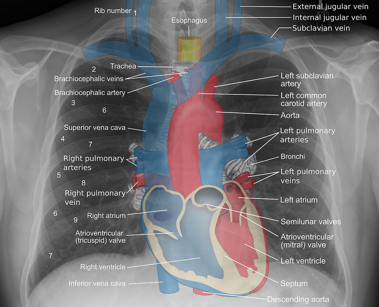
Cardiomediastinal anatomy on chest radiography | Image ... from images.radiopaedia.org Evaluation of a chest radiograph may appear to be simple, but is in fact a complex task requiring careful observation, sound understanding of chest anatomy, and knowledge of the principles of physiology and pathology. It is almost always the first imaging study ordered to evaluate for pathologies of the thorax, although further diagnostic imaging, laboratory tests. Radiographic anatomy of the chest and abdomen: Gillian lieberman forthe harvard 62. Both lungs should be well expanded and similar in volume. It first appears too complicated to read the chest xrays because we barely know what. We've labeled and outlined the main visible anatomical structures such as lungs. The interpretation of a chest film requires the understanding of basic principles.
The interpretation of a chest film requires the understanding of basic principles.
In this article we will focus on: Abcde aproach the anatomy of the heart can appear artificially larger due to this image orientation. It is almost always the first imaging study ordered to evaluate for pathologies of the thorax, although further diagnostic imaging, laboratory tests. A collection of anatomy notes covering the key anatomy concepts that medical students need to learn. Major structures are shown in fig. It first appears too complicated to read the chest xrays because we barely know what. Xray is a type of radiography and most widely used investigation. Airway (midline, patent) bone (eg, fractures, lytic lesions) cardiac silhouette size (diaphragm) edge (borders) of the heart (to rule out lingular and left middle lobe pneumonia or infiltrates). Chest radiographs are the most common film taken in medicine. In fact every radiologst should be an expert in chest film reading. Living anatomy of the chest for 1st year medical students original version compiled by dr. Heart and great vessels — assessment of the cardiovascular anatomy includes assessment of heart and chamber size as well as the position and size of the great. Gillian lieberman forthe harvard 62.
It is almost always the first imaging study ordered to evaluate for pathologies of the thorax, although further diagnostic imaging, laboratory tests. Conclusion of living anatomy of the chest congratulations! This imaging method can also check how a patient is responding to specific treatments. You have completed this module. The radiologist needs to know both the structures within the mediastinum forming the mediastinal margins and the.
File:Mediastinal structures on chest X-ray, annotated.jpg ... from upload.wikimedia.org The interpretation of a chest film requires the understanding of basic principles. Xray is a type of radiography and most widely used investigation. In this article we will focus on: Airway (midline, patent) bone (eg, fractures, lytic lesions) cardiac silhouette size (diaphragm) edge (borders) of the heart (to rule out lingular and left middle lobe pneumonia or infiltrates). Abcde aproach the anatomy of the heart can appear artificially larger due to this image orientation. The radiologist needs to know both the structures within the mediastinum forming the mediastinal margins and the. It first appears too complicated to read the chest xrays because we barely know what. Is there any inhaled foreign body?
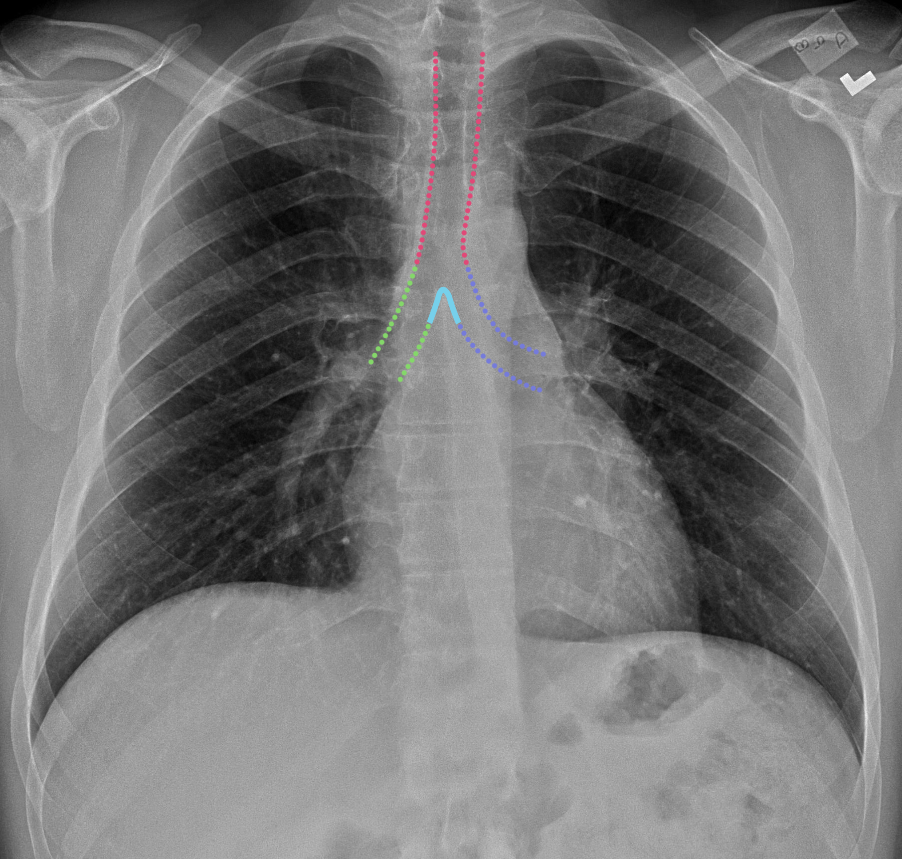
Look for lung and pleural pathology.
A method for examining a chest. It first appears too complicated to read the chest xrays because we barely know what. Major structures are shown in fig. In fact every radiologst should be an expert in chest film reading. L these two lobes are separated by a major fissure, identical to that seen on the right side, although often slightly more inferior in location. Living anatomy of the chest for 1st year medical students original version compiled by dr. Xray is a type of radiography and most widely used investigation. Gillian lieberman forthe harvard 62. A collection of anatomy notes covering the key anatomy concepts that medical students need to learn. Airway (midline, patent) bone (eg, fractures, lytic lesions) cardiac silhouette size (diaphragm) edge (borders) of the heart (to rule out lingular and left middle lobe pneumonia or infiltrates). In this article we will focus on: It is almost always the first imaging study ordered to evaluate for pathologies of the thorax, although further diagnostic imaging, laboratory tests. Is there any inhaled foreign body?
Air spaces normally seen in. It is almost always the first imaging study ordered to evaluate for pathologies of the thorax, although further diagnostic imaging, laboratory tests. The interpretation of a chest film requires the understanding of basic principles. A collection of anatomy notes covering the key anatomy concepts that medical students need to learn. Therefore, knowing the basics and pathologies in the ed setting is very important.
CaseStacks.com - Chest X-Ray Anatomy for Medical Students from www.casestacks.com Living anatomy of the chest for 1st year medical students original version compiled by dr. This imaging method can also check how a patient is responding to specific treatments. Each of these anatomical structures should be viewed using a systematic approach. Therefore, knowing the basics and pathologies in the ed setting is very important. Radiographic anatomy of the chest and abdomen: There are also important structures that are obscured or become visible. Common symptoms that can be diagnosed using chest. Major structures are shown in fig.
Conclusion of living anatomy of the chest congratulations!
Anatomy of a chest x ray. Therefore, knowing the basics and pathologies in the ed setting is very important. We've labeled and outlined the main visible anatomical structures such as lungs. Conclusion of living anatomy of the chest congratulations! Airway (midline, patent) bone (eg, fractures, lytic lesions) cardiac silhouette size (diaphragm) edge (borders) of the heart (to rule out lingular and left middle lobe pneumonia or infiltrates). Chest radiographs are the most common film taken in medicine. Common symptoms that can be diagnosed using chest. A method for examining a chest. L the portion of the left lung that corresponds anatomically to the right middle lobe is incorporated into the left upper lobe. Gillian lieberman forthe harvard 62. Xray is a type of radiography and most widely used investigation. Each of these anatomical structures should be viewed using a systematic approach. Radiographic anatomy of the chest and abdomen:
Therefore, knowing the basics and pathologies in the ed setting is very important anatomy of chest. Labeled chest radiographs teaching radiologic anatomy with a level of detail appropriate for medical students.


0 Komentar